MRI model trained on simulated photo data holds up on unseen scanners
A preprint trains an MRI reconstruction model mostly on fake scanner data made from everyday photographs. On scans from other hospitals and machines, it beat methods trained on real patient scans.

What the study shows
MRI scans are slow. One way to speed them up is to collect less raw data and let software fill in the gaps. This step is called reconstruction. AI reconstruction works well on scans like those it was trained on, but often fails on other scanners, hospitals or protocols.
The authors, from the Shenzhen Institutes of Advanced Technology, Peking University Shenzhen Hospital and Tencent, built a different training source. Their simulator takes ordinary photographs and turns them into fake raw MRI measurements. It follows the physics of how MRI receiver coils pick up signal. It adds realistic noise and invents a new coil layout for every example. The model, called SIM, is a vision transformer, a general-purpose image network, with up to about 1 billion parameters.
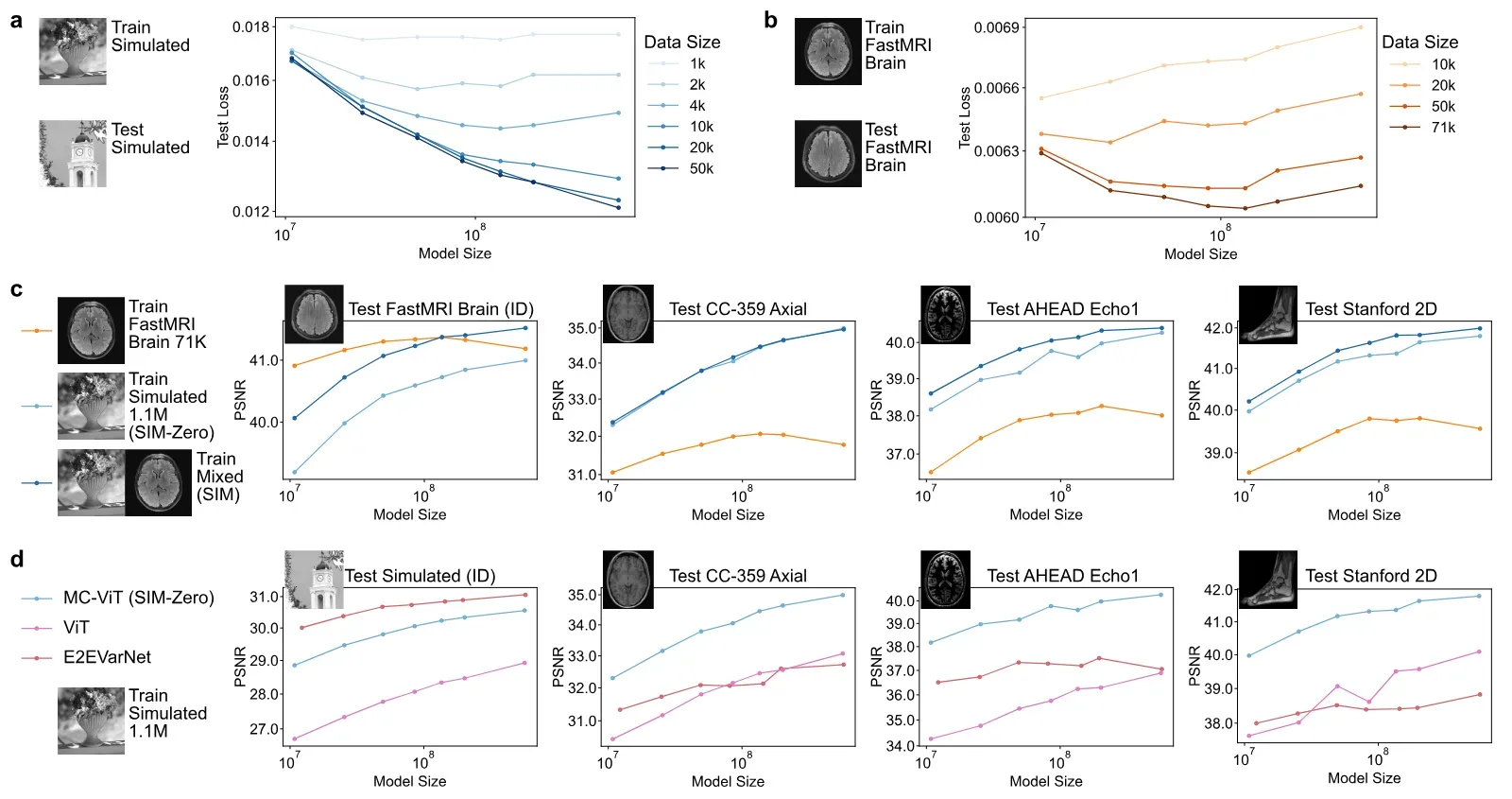
They tested it on 14 datasets: 8 public and 6 from their own institutions. These span five scanner makers, magnet strengths from very low to very high field, and many body parts. Baseline methods were trained on one large public brain dataset, fastMRI Brain. The authors report that SIM beat the second-best method on all 8 unseen public test sets. The margin was about 1 to 4 decibels on PSNR, a standard pixel-error score where higher is better.
A version trained only on simulated data did almost as well as the full model on unseen data. Adding real brain scans mainly helped on brain scans like the training set.
The authors also report that the model kept improving as it grew and saw more simulated data. Models trained on real brain scans alone stopped improving and overfitted, meaning they memorised training data instead of learning general rules.
In a blinded study, two radiologists rated brain scans from 50 people, half of them with Alzheimer's disease. At the mildest speed-up tested, every SIM scan was rated good enough for diagnosis. The main comparison method, E2EVarNet, produced no scans rated fully diagnostic at that speed. The study is a preprint and has not been peer reviewed.
Our reading
In our reading, the most useful finding is not the headline score. It is that a model trained without any patient scans came close to the full model on unfamiliar scanners. That points to the physics simulation, not the anatomy in the training data, doing most of the work. The authors' own ablation tests, where they remove one part at a time, support this. Removing simulator features hurt more than swapping the network design.
The comparison needs care. The rival methods were trained on one dataset from one hospital network. So SIM was up against models that had seen little variety. The paper does not test whether supervised models trained on varied real scans would close the gap. The large drop for those baselines on new scanners is real, but part of it reflects their narrow training.
We rate the work at level II, Benchmarked, on our four-level scale. It reports results on several public datasets against nine published methods. We found no released code or model weights in the paper, so it does not reach level III. A concrete use, if it holds up: a clinic with an unusual low-field scanner could shorten exams with a model never trained on its own scans.
Read the rest of this story
Membership is free. You get every article in full and the Issue every Sunday.
We send a sign-in link, no password needed. Already a member? Sign inReadiness assessment
| Level | Status | Evidence |
|---|---|---|
| I Lab only | Met | A working model with tested results on retrospective data, including a small reader study. |
| II Benchmarked | Met | Evaluated on 14 datasets, 8 of them public (fastMRI, CC-359, M4Raw, AHEAD, Stanford 2D and 3D). Compared with nine published methods. Results are on public datasets, though run by the authors and in a preprint. |
| III Open source | Not yet | The paper says simulation code would be distributed, but gives no link, repository or weights release. No code, weights or data link found in the text. |
| IV In production | Not yet | No documented deployment by a named organisation. The authors state that prospective multicentre studies are still needed. |
What would move it up
To reach level 3, release the simulation engine, model code and weights under a stated licence. To reach level 4, show prospective use on scanners at a named hospital or vendor, with regulatory status, and compare against locally trained or vendor reconstruction on accelerated scans. Independent replication on other sites and pathologies would also help.
Checked automatically every week, last on 16 November.
The study
Hong Shang, Zijian Zhan, Sen Jia et al.. Physics-based simulation unlocks scaling laws for generalizable MRI reconstruction. arXiv, 11 November 2026. Read the paper
Notes
- Shang H, Zhan Z, Jia S, et al. Physics-based simulation unlocks scaling laws for generalizable MRI reconstruction. arXiv 2611.15758, 11 November 2026. Preprint, not peer reviewed. CC BY 4.0. Source
- No code, weights or data link was found in the paper. The authors say simulation code would be distributed.
- Exact figures: at an effective 3.2x acceleration, SIM exceeded the second-best method by 1.06 to 3.95 dB PSNR and 0.008 to 0.045 SSIM across 8 out-of-distribution test sets. E2EVarNet's drop from in- to out-of-distribution reached 10.01 dB PSNR; SIM's maximum drop was 5.64 dB.
- Training: about 1.01 to 1.1 million simulated samples from the SA-1B image set; final models also used 70,748 real fastMRI Brain slices. Main models had 510 million and 1.01 billion parameters.
- Reader study: 50 subjects (25 controls, 25 Alzheimer's patients), 1,050 volumes, two radiologists. At 5.1x, non-diagnostic rates were 4%, 0% and 1% for SIM (T1, T2, FLAIR) against 100%, 66% and 99% for E2EVarNet. Weighted kappa 0.91 for image quality and 0.73 for usability (0.51 for T2 usability).
- Downstream: T2* mapping error reduced by at least 1.83 ms against E2EVarNet. CEST recovery constants 282 s and 432 s against 279 s and 429 s for the reference, from one subject at 5.1x.
- Readiness: level II, Benchmarked. Public benchmark results reported; no released code or weights; no documented production use.
Edited and signed by
Stefan PerdalEditor and publisher86 stories editedStefan read the study, set the readiness level and approved this story. An AI system found the paper and wrote the first draft.
How this was made
An AI model read the study and wrote this article in the structure Science Underway uses. An editor reviewed it and approved it for publication. Check the original study before you rely on the details.
How we workNo corrections